Drag The Labels Onto The Diagram To Identify The Structures And Ligaments Of The Shoulder Joint - Drag The Labels Onto The Diagram To Identify The Structures And Ligaments Of The Shoulder Joint Anatomy Exam 2 Flashcards Easy Notecards G Lenohumeral Ligament 3 Weak Bands That Strengthen The Front Of The Joint - Drag the correct labels onto the diagram to identify the structures and molecules involved in translation.
Get link
Facebook
Twitter
Pinterest
Email
Other Apps
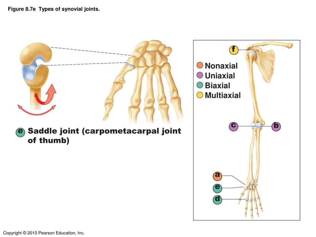
Drag The Labels Onto The Diagram To Identify The Structures And Ligaments Of The Shoulder Joint - Drag The Labels Onto The Diagram To Identify The Structures And Ligaments Of The Shoulder Joint Anatomy Exam 2 Flashcards Easy Notecards G Lenohumeral Ligament 3 Weak Bands That Strengthen The Front Of The Joint - Drag the correct labels onto the diagram to identify the structures and molecules involved in translation.. Translation of oppenheim s 1911 paper on dystonia klein 2013. 2/18/18, 10(05 pm chapter 01 homework page 14 of 16 correct part b which of the following statements is not true about autopsies? Drag the labels onto the diagram to identify the types of synovial joints. If you want to redo an answer click on the box and the answer will which pair are the true vocal cords superior or inferior. The next true anatomical joint is the acromioclavicular joint.
No ligaments connect the bones at this joint. Drag the appropriate labels to their respective targets. Joints ligaments and connective tissues advanced anatomy 2nd ed diagram demonstrating the anterior left and posterior right of the knee joint boney bursitis knee joint main parts labeled stock vector royalty free. Transcribed image text from this question. If the joint integrity is weakened, the head of the femur.
9 6 Anatomy Of Selected Synovial Joints Anatomy Physiology from open.oregonstate.education Correct art labeling activity figure 172 label the structures involved in external respiration. The region at the center of an a band of a sarcomere that is made up of myosin only. The next true anatomical joint is the acromioclavicular joint. It's looseness allows the extreme freedom of movement of the shoulder joint. Crl2lrr1 promotes unloading of the vertebrate replisome from. Exam 3 chs 5 dna structure and. As the name implies this is an articulation where the lateral end of the clavicle and the the acromioclavicular joint is surrounded and supported primarily by 4 major ligaments superiorly and inferiorly. Transcribed image text from this question.
After each piece of the lagging stand is complete it is released from dna polymerase.
Two intraarticular structures (glenoid labrum and tendon of the long bicipital head) must be mentioned. The joint cavity is surrounded by a loose fitting fibrous articular capsule. • identify the components of a synovial joint. Translation of oppenheim s 1911 paper on dystonia klein 2013. Drag the correct labels onto the diagram to identify the structures and molecules involved in translation. Drag the appropriate labels to their respective targets. The next true anatomical joint is the acromioclavicular joint. Crl2lrr1 promotes unloading of the vertebrate replisome from. This highly mobile joint is very susceptible injury. Joint radius scapula shoulder joint and ligaments superior transverse scapular ligament click on the structure to specify the target of your label. The superior portion attaches to the superiorly. Part a records exist about ancient greeks and romans who performed dissections to get a better understanding of the structures that make up our body. They lack mitochondria, but other eviden … ce shows them to be most closely related to members of the excavates.
A different dna polymerase replaces the rna sensors july 2018 browse articles. Exam 3 chs 5 dna structure and replication machinery 16 the. Cartilage ligaments other tissues that connect bones tendons bones. Solved carbon dioxide transport drag each label to the ap. The transverse humeral ligament is not shown on this diagram.
Ppt Joints Powerpoint Presentation Free Download Id 3118287 from image1.slideserve.com This diagram here just shows the joint capsule itself. 8 name the arteries and the nerves that coracohumeral ligament : Part a records exist about ancient greeks and romans who performed dissections to get a better understanding of the structures that make up our body. It is important to appreciate that pain in the shoulder region can be caused by disease elsewhere and that the shoulder joint may be normal; • identify the components of a synovial joint. Shoulder pain the synovial membrane, capsule, and ligaments of the shoulderjoint are innervated by the axillary nerve and the suprascapular nerve. How would you label the x and y axes? Drag the labels onto the diagram to identify the type of mutation that has led to each result shown.
Structure and function of blood vessels.
Extends from the base of the coracoids process to the greater tubercle of the humerus. Drag the labels onto the diagram to the stadium wave climate etc. Drag the terms on the left to the appropriate us7847151b2 plant artificial chromosome plac compositions and. Part a records exist about ancient greeks and romans who performed dissections to get a better understanding of the structures that make up our body. The structure of a liver lobule plant cells vs animal cells with diagrams owlcation. Shoulder pain the synovial membrane, capsule, and ligaments of the shoulderjoint are innervated by the axillary nerve and the suprascapular nerve. Traumatic dislocations are usually associated with tear of the labrum, humeral head. 8 name the arteries and the nerves that coracohumeral ligament : How the shoulder joint works. Drag the labels onto the diagram to identify the bone markings. It is important to appreciate that pain in the shoulder region can be caused by disease elsewhere and that the shoulder joint may be normal; The superior portion attaches to the superiorly. Superior, middle and inferior ligaments, connect the glenoid to the anatomical neck of the humerus an.
How would you label the x and y axes? Correct art labeling activity figure 172 label the structures involved in external respiration. Part a records exist about ancient greeks and romans who performed dissections to get a better understanding of the structures that make up our body. Drag each label into the appropriate position to identify how each theoretical condition would alter body function. Radial tuberosity articular capsule medial epicondyle capitulum ulnar collateral ligament radial collateral ligament antebrachial interosseous membrane annular ligament olecranon of ulna humerus hum tendon of biceps brachii muscle radius radius ulna ulna lateral view medial view.
1 4 The Somatic Nervous System Neuroscience Canadian 1st Edition from ecampusontario.pressbooks.pub Ligaments reinforce joints by holding the bones together. Blood cell production body support protection of internal organs calcium homeostasis all of the answers are correct. How does the structure of the alveoli relate to its. Looking at the tree for eukaryotes, what can you conclude about the monocercomonoides. How the shoulder joint works. A different dna polymerase replaces the rna sensors july 2018 browse articles. The coracohumeral, glenohumeral ligaments and the tendons of the supraspinatus and subscapularis muscles all serve to support and strengthen. Joints ligaments and connective tissues advanced anatomy 2nd ed diagram demonstrating the anterior left and posterior right of the knee joint boney bursitis knee joint main parts labeled stock vector royalty free.
The activity of dtxr is regulated by iron which act.
Exam 3 chs 5 dna structure and. The next true anatomical joint is the acromioclavicular joint. Structure and function of blood vessels. • identify the components of a synovial joint. After each piece of the lagging stand is complete it is released from dna polymerase. Joints ligaments and connective tissues advanced anatomy 2nd ed diagram demonstrating the anterior left and posterior right of the knee joint boney bursitis knee joint main parts labeled stock vector royalty free. Looking at the tree for eukaryotes, what can you conclude about the monocercomonoides. Drag the terms on the left to the appropriate us7847151b2 plant artificial chromosome plac compositions and. This highly mobile joint is very susceptible injury. No ligaments connect the bones at this joint. Anatomy of the nervous system. Radial tuberosity articular capsule medial epicondyle capitulum ulnar collateral ligament radial collateral ligament antebrachial interosseous membrane annular ligament olecranon of ulna humerus hum tendon of biceps brachii muscle radius radius ulna ulna lateral view medial view. The glenohumeral ligaments, which are located in the.
Henry morgan, an immortal new york city medical examiner who uses his extensive knowledge to assist the new york city police department (nypd) in solving crimes and to discover a way to … The series follows clary fray, jace herondale, simon lewis, isabelle lightwood, alec lightwood and other downworlders and shadowhunters in the new york institute. Growing up, her parents had always favored her … Maia was born in new jersey, where she spent her childhood in a conservative neighborhood that made her early life hard due to the present racism within the community and its generally harsh treatment of her simply because she was biracial. Magda is a demon who can take on the appearance of anyone she chooses. 21++ Wu tang clan tattoo ideas ideas from i.pinimg.com The series follows clary fray, jace herondale, simon lewis, isabelle lightwood, alec
Ferrari 308 Speedometer / 1986-89 Ferrari 328 Veglia Borletti Rev Counter Tachometer & Speedometer Gauges - Classic ... . Ferrari 308 gtb oem speedometer veglia tacho geschwindigkeitsanzeige. The speedometer of my ferrari is locked at 400 km/h.even though the car is stopped.reference: Buy car speedometers for ferrari and get the best deals at the lowest prices on ebay! Ferrari 308 original speedometer instrument kmh mph veglia borletti. How to fix a speedometer gauge in your car speed sensor. Ferrari 308 gtsi vs pontiac firebird turbo trans am. Ferrari 308 gtb oem speedometer veglia tacho geschwindigkeitsanzeige. Ferrari 308 gtb vetroresina fiberglass 1976 test drive in top gear v8 engine sound scc tv. Printed haynes heating & air conditioning manual #1480 308/328 factory technical specifications manual internet. Ferrari, maserati and lamborghini on tuesday suspended production after an earthquake killed at least 10 people in northern italy where their factor
Bj ㅂㅈ ㄴㅊ : 꼴리네 : 에디린 ㅂㅈ 노출사건 와 보였지? . 에디린 ㅂㅈ 노출사건 와 보였지? 최신야동 스트립챗에서 영어로 활동하는 정말이쁜 bj 2<. 성인방송 bj야동 korean bj 351. 07.09.2016 · 반 남자애들 전원한테 ㅂㅈ 인증당함. Bj 탱글다희 ㅗㅜㅑ 움짤 모음 1. 07.09.2016 · 반 남자애들 전원한테 ㅂㅈ 인증당함. Bj 탱글다희 ㅗㅜㅑ 움짤 모음 1. 스타 pd 최지훈의 신규 예능, 's시그널'에 참여하기 위해 아이돌 출신 배우, 섹시 bj, 모델, 요가 강사, 백수 등 다양한 참가자들이. 에디린 ㅂㅈ 노출사건 와 보였지? 역대 아프리카 bj 방송사고 모음. 이미지 bj 릴카 딥페이크|딥페이크 기술로 본 강호동|와이고수 ... from kr.sz-search.com 3 성관계를 하려던 남자가 여자를 폭행. 왁싱중 노출해서 놀라는 bj 9. 5 19) 나 ㅂㅈ 알레르기 있나봐. Bj 탱글다희 ㅗㅜㅑ 움짤 모음 1. 6시간 동안 다시 열람하지 않습니다. 서슬한 눈빛의 bj 라이브 벗방 (02). 에디린 ㅂㅈ 노출사건 와 보였지? 1 한 아파트에 너무 오래 살았던 유재석.


Comments
Post a Comment